Prostate biopsy is a procedure in which pathological tissue is taken to detect the presence or absence of prostate cancer in patients with elevated blood tests (PSA) or abnormalities in digital rectal examination or significant lesions detected in multiparametric prostate MRI.
In traditional systematic prostate biopsy, tissue pieces are taken from 12 quadrants of the prostate and examined under ultrasound guidance.
Since the ultrasonography (USG) used during the biopsy procedure has a low sensitivity in detecting the cancer focus within the prostate, especially low-stage prostate cancers may not be diagnosed.
With the help of technological developments in recent years, the cancer focus within the prostate has begun to be detected much better with multiparametric MRI.
With the help of MRI applied to the patient before prostate biopsy, the rate of detection of prostate cancer by experienced urologists increases. Because these MRI images are transferred to ultrasonography and show us the right path.
MRI fusion biopsy procedure; It is a method that allows us to take a biopsy from the right place by transferring the images taken before prostate biopsy to ultrasonography. It is a navigation system. It protects us from unnecessary biopsy and provides us correct diagnosis.
The most superior method for imaging lesions that are a sign of prostate cancer is technically multiparametric MRI, in which different sequences are taken in a single session. With the 3 TESLA MRI technology at Doruk Nilüfer Hospital, multiparametric prostate MRI is evaluated at the highest level. If there is a suspicious lesion (PIRADS-3,4,5),MRI fusion prostate biopsy is performed.
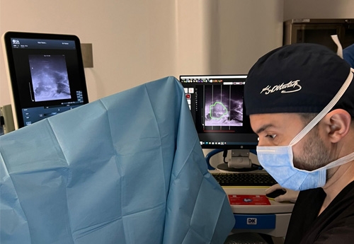
Before MRI fusion prostate biopsy, multiparametric MRI is applied to the patient to detect a suspicious lesion located in the prostate. During the procedure, the ultrasonography device that images the prostatic tissues is guided by the lesion that is suspicious for cancer seen on MRI with the help of the fusion device. It is beneficial to use antibiotics and enemas to prevent infections before the procedure.
Due to this smart prostate biopsy system, the probability of diagnosing cancerous tissues increases to a very high level. Considering the anxiety of our patients during prostate biopsy, it also saves patients from repeated unnecessary biopsies. The possibility of misdiagnosis of existing prostate cancer is minimized, ensuring that patients receive more accurate treatment.
It is performed with the help of a previously taken MRI image in a patient suspected of having prostate cancer and devices that transfer these images to ultrasonography and make markings. Considering the pain of the procedure and the anxiety of the patients, prostate biopsy is a procedure that should be performed under general or regional anesthesia in operating room conditions.
The samples to be taken are taken with the help of a biopsy needle, but this sampling process has two sub-methods in MR fusion biopsy: transrectal and transperineal. In other words, MRI fusion biopsy is divided into two.
The transrectal method is the removal of prostate tissues by entering through the anus, and it has the advantage of being easy to apply. However, since this method is entered through the anus, the risk of serious infection is much higher than the transperineal method.
Transperineal access is the removal of prostate tissues by inserting a needle through the skin just above the anus. A sample is not taken from the anus. Since the biopsy is taken from a clean area, the possibility of infection is less than the transrectal method and the ease of application with the MRI Fusion device makes this technique the gold standard. This is the method mainly recommended by the European Association of Urology.
MRI fusion prostate biopsy is a method that can be performed painlessly when performed under anesthesia, and the possibility of infection is very low, especially if access is made via transperineal route. It is the best method in the diagnosis of prostate disease (benign prostate hyperplasia and prostate cancer).
Although the prostate biopsy procedure takes approximately 15-20 minutes, the process may take up to approximately 45 minutes due to preliminary preparation and anesthesia.
This procedure was performed by Assoc. Prof. Dr Arif Demirbaş at Doruk Hospitals Turkey. It is performed painlessly and comfortably under anesthesia.
It is important for the pathology result to be obtained early, both for the patient's concern about the result and for the planning of the treatment. At Bursa Doruk Hospitals, pathology results are reported on average in 5-7 days.
MR fusion biopsy prices vary depending on hospital conditions, the anesthesia method used, the quality and operating system of the MR Fusion device used, and the experience of the performing physician.
Whatsapp: +90 532 485 0016
Instagram: @drarifdemirbas
Youtube: @drarifdemirbas
Note: The page contents are for informational purposes only and a doctor's consultation is required for your diagnosis and treatment.
Hello how much yo you charge to do prostate byops
Hello. We perform transperineal Mr fusion biopsy. We will send you the details.
 Assoc. Prof. Dr. Arif DemirbasUrology Specialist
- I will definitely recommend this hospital in the future.
- I cannot recommend Dr. Arif Demirbaş enough.
- God continue to bless you in all your good work.
All TestimonialsMeet Alpha from Sierra Leone who just undergone a PCNL and Utereoscopy surgeries at the Doruk hospital in Bursa. I want to start by sending a massive thank you to Dr Arif for exhibiting such a {...}
24.01.2025I cannot recommend Dr. Arif Demirbaş enough. My father had been living with a catheter for over two years due to benign prostatic hyperplasia, which also caused bladder stones and an inguinal h{...}
24.01.2025Thanks to you Dr Arif for your successful kidney stone surgery on my Son Ryan since after we came back from Turkey to date Alhamdulillah Ryan is very ok Alhamdulillah bravo for your Good work. {...}
24.01.2025